
Reconstructive Plastic Surgery
Skin Cancer and Mole Removal
Skin moles, or naevi, may be present from birth or develop later in life. They vary widely in colour, size, shape and texture and should be assessed if they change.
Their development over time may be influenced by internal factors, such as hormones, and external factors, including the environment and sun exposure. Most are benign skin lesions whose course is monitored for any changes.
In a small proportion of cases, however, malignant lesions such as melanoma may develop. People with moles should protect their skin from sun exposure and avoid injuring the moles.
Any change in the colour, size, shape or texture of a mole should prompt assessment by a dermatologist.
In a small proportion of cases, however, malignant lesions such as melanoma may develop. People with moles should protect their skin from sun exposure and avoid injuring the moles.
Any change in the colour, size, shape or texture of a mole should prompt assessment by a dermatologist.
Interview with Dr Andreas Moutsoudis for cityportal.gr on Skin Cancer Prevention and Early Diagnosis
People with numerous skin moles, particularly pigmented naevi, should be examined by a dermatologist once or twice a year. Digital mole mapping enables detailed monitoring over time.
Skin tumours include benign lesions, such as papillomas and cysts, as well as skin cancers, principally basal cell carcinoma, squamous cell carcinoma and melanoma. Environmental factors, sun exposure, skin type and genetic predisposition may contribute to the development of skin cancer.
Early diagnosis and treatment are important for the appropriate management of skin cancer. A skin lesion that does not resolve, or a pre-existing lesion that changes, should therefore be assessed promptly by a specialist dermatologist.
Skin tumours include benign lesions, such as papillomas and cysts, as well as skin cancers, principally basal cell carcinoma, squamous cell carcinoma and melanoma. Environmental factors, sun exposure, skin type and genetic predisposition may contribute to the development of skin cancer.
Early diagnosis and treatment are important for the appropriate management of skin cancer. A skin lesion that does not resolve, or a pre-existing lesion that changes, should therefore be assessed promptly by a specialist dermatologist.
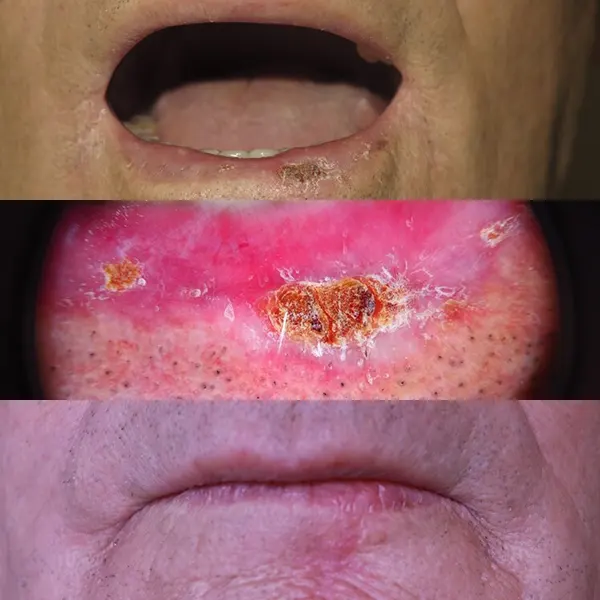
Squamous Cell Carcinoma of the Lip
Squamous cell carcinoma is the second most common skin cancer, and the lips are a frequent site of the disease. Its early form is known as actinic cheilitis, and early diagnosis and treatment contribute to effective management. When the tumour invades more deeply, surgical excision and biopsy, together with staging of the disease, are required.
The case presented involved an 83-year-old patient who underwent wide excision and reconstruction with a local flap. The patient has remained free of disease and continues regular follow-up with a dermatologist and plastic surgeon to support early recognition of any new lesions.
Functional outcomes relating to chewing and speech, as well as the aesthetic outcome, were assessed as satisfactory in this individual case.
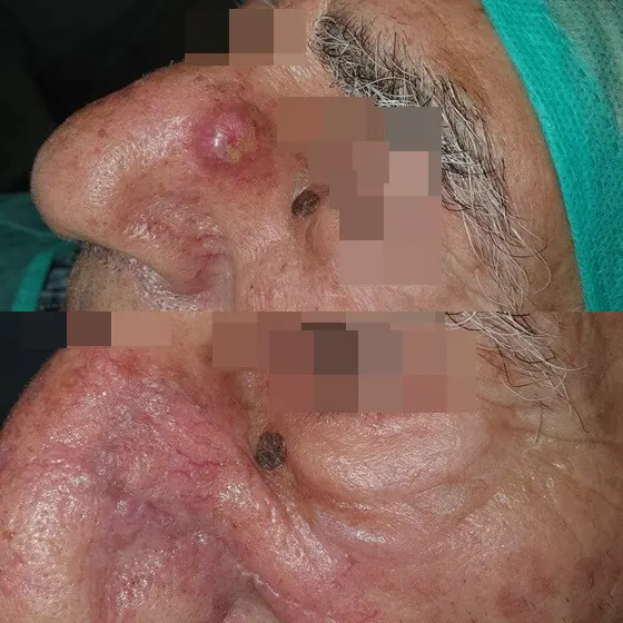
Squamous Cell Carcinoma of the Nose
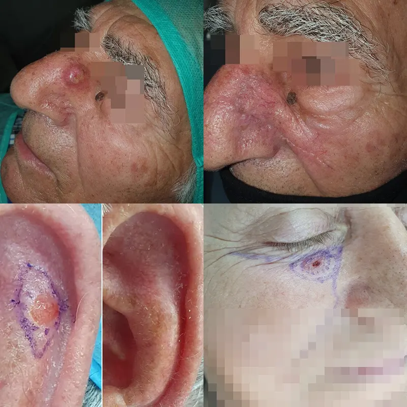
This is the second most common skin cancer and occurs after the age of 50. Early assessment with dermoscopy and biopsy confirms the presence of neoplastic cells, and the proposed treatment is surgical excision with clear margins in accordance with the relevant protocol.
The case presented involved a 75-year-old patient who underwent surgery under local anaesthesia. The resulting defect was reconstructed with a V-Y advancement flap.
The postoperative course progressed without complications, and the functional and aesthetic outcomes were assessed in this individual case. The patient will continue regular follow-up for early recognition of any new lesions.
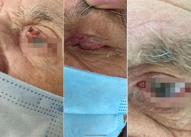
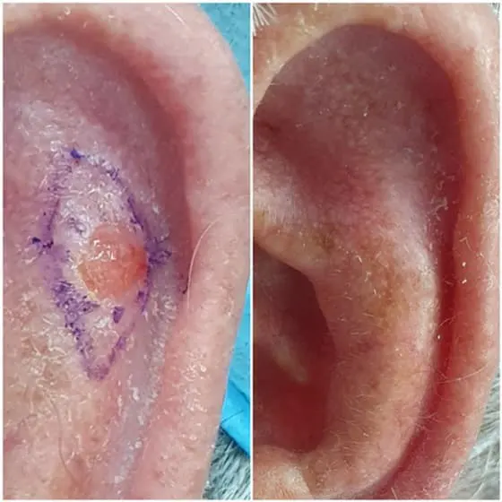
Basal Cell Carcinoma of the Ear
Direct closure was performed, and the aesthetic outcome was assessed postoperatively.
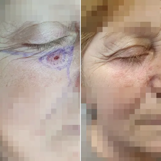
Basal Cell Carcinoma of the Upper Eyelid
This basal cell carcinoma of the upper eyelid involved the full thickness of the skin and the conjunctiva. Reconstruction required a particularly demanding procedure and detailed knowledge of the anatomy and flap techniques used to transfer adjacent skin.
Biopsy showed that the carcinoma had been excised with clear margins. Upper-eyelid reconstruction produced a functional outcome that continues to be assessed during follow-up. The patient will continue regular monitoring with dermoscopy.
Another case involved basal cell carcinoma of the lower eyelid. It was treated with a cheek advancement flap. Skin reconstruction and healing are assessed during regular follow-up.
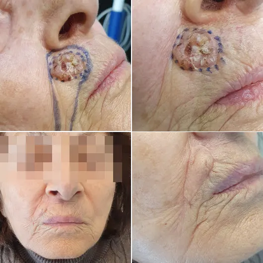
The following case involved basal cell carcinoma of the upper lip, treated by surgical excision under local anaesthesia in an outpatient setting.
The dermoscopic appearance of the basal cell carcinoma was an indication for its removal. The defect was reconstructed with a V-Y advancement flap, and healing at three months was considered satisfactory from both functional and aesthetic perspectives.
The patient continues dermatological follow-up twice a year to support prevention or early diagnosis of precancerous or cancerous lesions.
The following case involved squamous cell carcinoma arising in an area of actinic cheilitis in an 82-year-old patient. Its characteristic dermoscopic appearance required surgical excision and reconstruction of the resulting wound.
Three local flap options were considered for reconstruction of the defect after surgery. Histology was consistent with the dermoscopic findings, and complete removal of the lesion was recorded. The patient can eat normally and her speech was not affected.
The postoperative photograph was taken two months after surgery. The patient will continue follow-up three times a year.
The following case involved basal cell carcinoma of the patient's left temple. Reconstruction was performed with a rhomboid flap under local anaesthesia.
The carcinoma was treated under local anaesthesia and excised with clear margins. Functional and aesthetic outcomes were assessed as satisfactory in this individual case.
The patient followed the postoperative instructions, and at six months there was no evidence of recurrence.
The final case shown is a squamous cell carcinoma located at the nasal tip.
Frequently Asked Questions
How is a skin mole managed?
Moles are assessed for changes in colour, size, shape or texture. Depending on the findings, monitoring or surgical removal under local anaesthesia may be recommended.
Which changes in a mole require assessment?
Any change in a mole’s shape, colour, size or texture requires attention. Further development, swelling or bleeding should be assessed by a specialist dermatologist.
How is digital mole mapping performed?
Digital mole mapping uses detailed photographic records to monitor moles over time, usually once or twice a year.
What does mole removal involve?
Removal is performed under local anaesthesia through a small incision or biopsy. Healing and the resulting scar vary according to the lesion, its location and the individual course of recovery.
What should be monitored in relation to skin cancer?
Basal cell and squamous cell carcinomas occur more frequently after the age of 50 and in areas with substantial sun exposure. Early assessment using dermoscopy and biopsy is important for planning appropriate treatment.
How often should moles be examined?
Examination once or twice a year may be recommended, particularly for people with numerous moles and especially pigmented naevi, according to the dermatologist’s assessment.
What does surgical removal of a skin tumour involve?
The procedure includes biopsy and excision of the tumour with clear margins. Reconstruction may use direct closure or a flap, depending on the anatomical site.
What aesthetic outcome can be expected after mole removal?
The aesthetic outcome and the appearance of the scar depend on the lesion, its location, the surgical technique and the individual healing response. Medical follow-up forms part of postoperative care.
What is the difference between basal cell and squamous cell carcinoma?
Basal cell carcinoma is more common and usually grows slowly. Squamous cell carcinoma may occur on areas such as the lips or nose and requires prompt surgical assessment.
How are moles and skin tumours assessed and treated?
Assessment may include dermoscopy and digital mole mapping. When surgery is indicated, excision and reconstruction are planned by the relevant dermatology and plastic-surgery specialists.
Would you like to discuss which option is suitable for you?
The right indication and a personalised treatment plan are determined after medical assessment.